What is the anterior cruciate ligament?
The Anterior Cruciate Ligament (ACL) is one of the four major ligaments in the knee that connects the femur (thigh bone) to the tibia (shin bone). It plays a crucial role in maintaining knee stability, especially during activities that involve sudden stops, jumps, and changes in direction. The ACL is positioned in the centre of the knee and prevents the tibia from sliding out in front of the femur, while also providing rotational stability.
ACL injuries are among the most common knee injuries, particularly in sports and physical activities that involve a high degree of movement complexity and force. Understanding the ACL’s role and the nature of its injuries is essential for both prevention and treatment.
Anatomy of the ACL
The ACL is located deep within the knee joint, crossing diagonally in front of the Posterior Cruciate Ligament (PCL), forming an “X” shape that provides crucial stability to the knee during movement. It originates from the medial aspect of the lateral femoral condyle and runs inferiorly, medially and anteriorly and inserts in to the intercondylar eminence of the tibia.
The primary function of the ACL is to prevent the tibia from sliding too far forward in relation to the femur. It also provides rotational stability, preventing the knee from twisting abnormally during pivoting movements. This ligament is essential for activities that involve sudden stops, changes in direction, or jumping, making it particularly important in sports like soccer, basketball, and skiing.
The ACL is a vital component of the knee’s overall stability, working in conjunction with other ligaments, tendons, and muscles to maintain proper alignment and function. Alongside the PCL, medial collateral ligament (MCL), and lateral collateral ligament (LCL), the ACL ensures that the knee can move through its full range of motion without giving way or collapsing under stress.
Muscles surrounding the knee, particularly the quadriceps and hamstrings, also play a critical role in supporting the ACL. The quadriceps muscle group extends the knee, while the hamstrings work to flex the knee and help stabilize the joint by counteracting forces that could stress the ACL. Strong, balanced muscles can reduce the risk of ACL injury by absorbing some of the forces that would otherwise be transferred to the ligament.
What causes an ACL injury?
Like any ligament injury, ACL injuries typically occur when the knee is subjected to forces that exceed the ligament’s capacity to maintain stability. There are several common mechanisms through which ACL injuries can happen:
- Sudden Stops and Changes in Direction: One of the most frequent causes of ACL injuries is a rapid deceleration followed by a change in direction. This movement places a significant strain on the ACL as it works to stabilise the knee joint. Sports like rugby, basketball and football, which involve frequent cutting and pivoting, are particularly high-risk.
- Jumping and Landing: Improper landing technique after a jump is another common cause of ACL injuries. If an athlete lands with their knee extended or in an awkward position, the force can be transmitted through the knee in a way that overstresses the ACL, leading to a tear. This is often seen in sports like volleyball, basketball, football and gymnastics.
- Direct Impact: While less common than non-contact injuries, a direct blow to the knee, such as in a football or rugby tackle, can also cause the ACL to tear. This typically happens when the knee is struck from the outside, causing it to bend inward and rotate, overwhelming the ACL.
Common symptoms of an ACL tear
ACL injuries often occur suddenly and can be accompanied by a range of symptoms. Recognising these symptoms is crucial for prompt diagnosis and treatment. Common signs of an ACL injury include:
- A “Pop” Sound: Many individuals report hearing or feeling a “pop” in the knee at the moment of injury. This sound is often a clear indication that the ACL has been torn.
- Immediate Pain: A torn ACL typically causes significant pain almost immediately. The pain is usually intense and located in the center or back of the knee.
- Swelling: Swelling of the knee usually begins within a few hours of the injury. This swelling is due to bleeding within the joint and can cause the knee to become stiff and difficult to move very quickly.
- Instability: After an ACL injury, the knee may feel unstable or “give out,” especially during activities that involve turning. This instability is due to the loss of the ACL’s stabilising function.
- Limited Range of Motion: The combination of pain, swelling, and instability often results in a decreased range of motion in the knee. Individuals will often find it difficult to fully straighten or bend the knee.
- Difficulty Bearing Weight: Walking or putting weight on the injured leg can be painful and difficult, often leading to a limp or an inability to walk without assistance.
Diagnosis
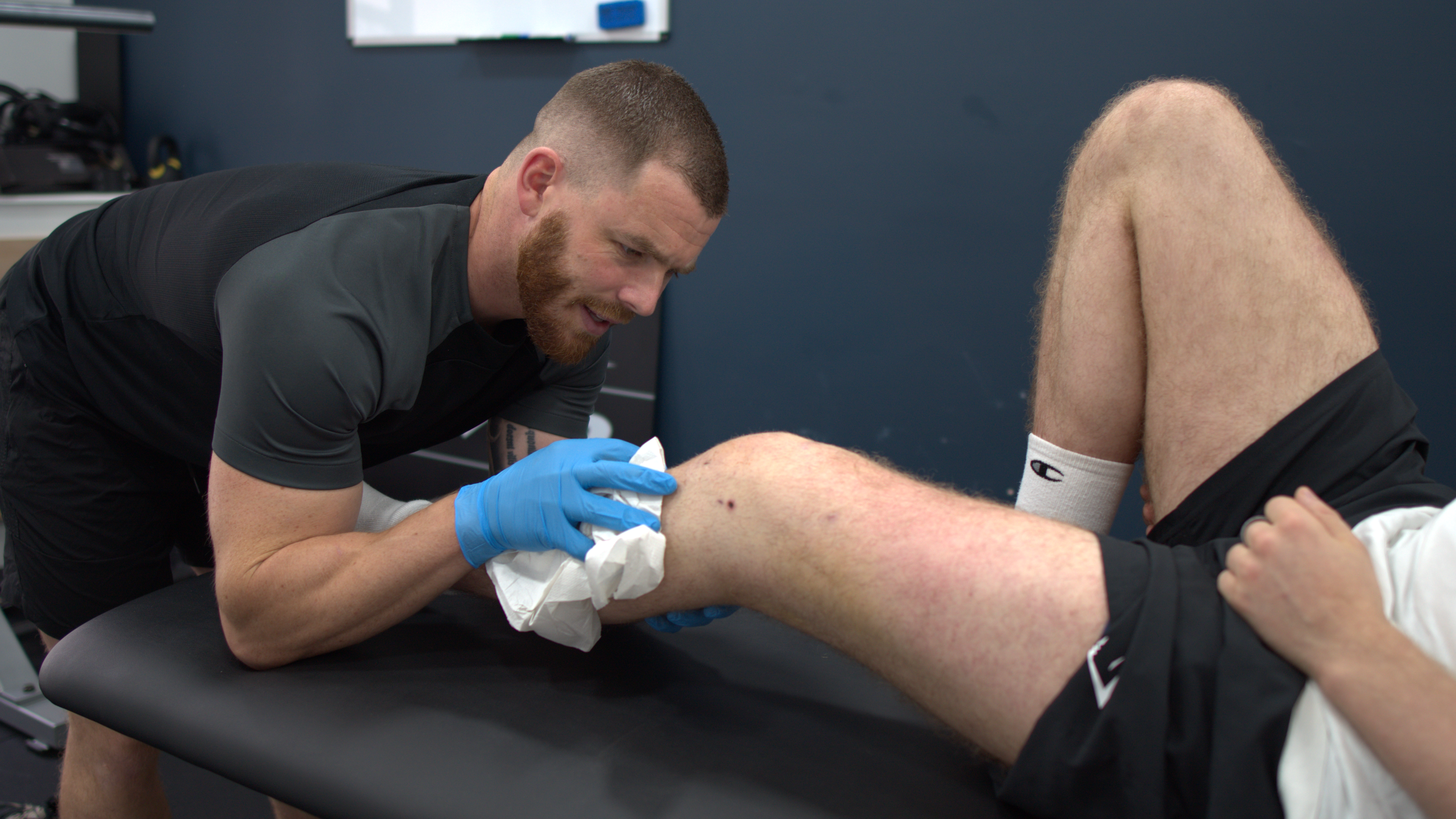
Diagnosing an ACL injury typically involves a combination of a physical examination and imaging to confirm the extent of the damage.
Physical Examination: A thorough physical examination by a physiotherapist is the first step in diagnosing an ACL injury. They will assess the knee’s stability, range of motion, and check for any signs of injury to other knee structures. Special tests like the Lachman test, anterior drawer test, and pivot shift test are used during this examination.
Magnetic Resonance Imaging (MRI): MRI is the gold standard for diagnosing ACL tears. It provides detailed images of both soft tissues and bone, allowing for a clear view of the ACL and any associated injuries, such as meniscal tears or damage to other ligaments. An MRI can also reveal partial tears or complete ruptures of the ACL.
X-rays: While X-rays do not show soft tissues like the ACL, they are often used to rule out fractures or other bone-related injuries that may have occurred simultaneously with the ACL injury. X-rays are particularly useful in cases of contact injuries where a fracture is suspected.
Arthroscopy: In some cases, particularly when the diagnosis is unclear or other knee injuries are suspected, arthroscopy may be used. This minimally invasive procedure involves inserting a small camera into the knee joint to directly visualize the ACL and other structures. Arthroscopy is also sometimes used to treat ACL injuries surgically.
Prompt and accurate diagnosis of an ACL injury is essential for determining the appropriate treatment plan. Whether surgical or non-surgical, early intervention can help prevent further damage and improve the chances of a full recovery.
Partial tears vs full ruptures
ACL injuries can vary in severity, ranging from mild sprains to complete tears. Understanding the difference between partial tears and complete ruptures is crucial for determining the appropriate treatment and rehabilitation approach.
- Partial Tears: A partial tear occurs when only some of the fibers of the ACL are torn, leaving the rest of the ligament intact. Partial tears can still cause pain, instability, and swelling, but the knee may retain some level of stability. These injuries may be treated conservatively with physiotherapy and bracing, although in some cases, surgery may be recommended if the instability is significant or if the individual is highly active in sports.
- Complete Ruptures: A complete rupture, or full tear, involves the tearing of all the fibers of the ACL, resulting in a complete loss of function of the ligament. In these cases, the knee is often highly unstable, and the individual may struggle to perform activities that involve pivoting, jumping, or sudden changes in direction. Complete ruptures almost always require surgical intervention, especially in athletes or active individuals, to restore stability and function to the knee.
What are osteochondral lesions?
Osteochondral lesions refer to areas where the cartilage that lines the ends of the bones in the knee (such as the femur or tibia) has been damaged along with the underlying subchondral bone. This can occur during an ACL injury, particularly when the knee experiences a high-impact force or twisting motion that not only tears the ligament but also crushes or shears off a piece of the bone and cartilage. These lesions can range from small, isolated areas of damage to larger, more complex injuries that affect significant portions of the joint surface.
Osteochondral lesions are a particularly serious problem because the knee’s articular cartilage does not heal as easily or as well as other tissues in the body. Cartilage lacks a direct blood supply, which limits its ability to regenerate. When an osteochondral lesion occurs, it can lead to persistent pain, swelling, and mechanical symptoms such as catching or locking of the knee. Over time, if left untreated, these lesions can result in joint degeneration, leading to early-onset osteoarthritis, a condition that can significantly impair mobility and quality of life.
Due to the complexity and potential severity of osteochondral lesions, they require prompt and expert attention. Diagnosis typically involves imaging studies such as MRI or CT scans, which can reveal the extent of the damage to both the cartilage and the underlying bone. Treatment may vary depending on the size and location of the lesion, as well as the patient’s activity level and overall knee stability. Options can range from conservative management, such as physical therapy and bracing, to surgical interventions.
Without appropriate treatment, osteochondral lesions can significantly impact long-term knee function. Even with successful ACL reconstruction, the presence of an untreated osteochondral lesion can lead to chronic knee pain, instability, and a decreased ability to participate in sports or other physical activities. The risk of developing post-traumatic osteoarthritis is also heightened, which can further complicate the recovery and rehabilitation process. Therefore, it is critical for anyone with an ACL injury to be thoroughly evaluated for the presence of osteochondral lesions and to receive specialised care from an orthopaedic expert experienced in managing complex knee injuries.
Recovering from an ACL injury can be a long and demanding journey, but with the right knowledge, treatment, and rehabilitation, many individuals can return to their pre-injury levels of activity. Rehabilitation is a vital component of this process, involving careful planning and execution of exercises, therapies, and lifestyle adjustments to ensure full recovery and minimise the risk of re-injury.
If you’re interested in diving deeper into the rehabilitation process, including detailed guidance on exercises, recovery timelines, and tips for a successful return to sports and daily activities, be sure to follow the next blog in this series. We’ll cover everything you need to know about navigating the road to recovery after an ACL injury.
The Sharp Physio Team
Listen to Jacks story…
Request A Free Discovery Call & Ask All The Questions You Need

Joe Sharp is the Clinical Director and Founder of Sharp Physiotherapy. With over 6 years of experience in private practice and a background working in professional football, Joe brings a practical, performance-driven approach to physiotherapy. As an HCPC-registered physiotherapist, he is passionate about delivering clear, effective treatment and helping patients stay active, mobile, and confident in their recovery.
Meet the Sharp Physiotherapy Team | Connect with Joe on LinkedIn


